


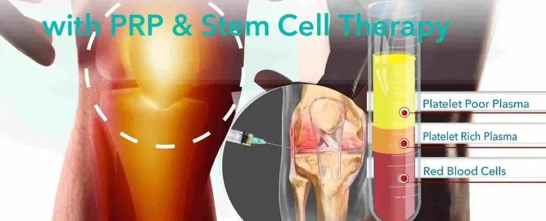
The half moon board is a fibrous cartilage located on the inside and outside joints of the tibial platform. The various opposite sex and unevenness of the biomechanics can meet various mechanics requirements of the knee joint, such as load bearing, maintaining knee coordination, stable exercise, and absorbing shocks. If the half moon plate injury cannot be treated in time, it will often cause osteoarthritis, and the main cause of the patient’s consultation is that the pain is aggravated and dysfunction. The half moon board can be divided into the following three areas, namely the white area, the red area and the red and white border area. There is no blood vessel distribution in the white zone, and local blood supply is not provided. Once damage is difficult to complete tissue repair. Therefore, it is difficult to repair after the half moon board injury, and most patients have poor prognosis. In recent years, with the continuous development of medical technology, the biochemical methods that promote the repair of half monthly dispenation have been widely used in the clinic and showed potential advantages. Plateau plasma plasma (PRP) can enhance the ability of semi -moon plate fibrocytocytes and white areas of cartilage cells to promote tissue healing and achieve significant results.
The characteristics of half moon board damage
1) The anatomy and function of the knee half moon board
As a fiber cartilage plate, the half moon board is between the tibial platform and the femoral cricket in the knee joint. The appearance characteristics of the half moon board are as follows: C -shaped inner side and O -shaped outside; the upper surface is sunken, the lower surface is flat; Connect. In addition, the half moon board can be attached to the edge of the tibial platform with the help of the outer coronary ligament and connect to the surrounding knee capsule, while the router tendon can pass through the outer and joint capsules of the half moon plate. Because the half moon plate blood supply is only provided by the surrounding tissue, once the surrounding tissue is damaged, the half moon plate is prone to necrosis, which will affect the knee function.
2) The injury mechanism of the knee half moon board
Adult knee joint half moon board can cause damage due to many external factors such as age, occupation and work intensity. Patients with young people are often torn, while senile patients are often closely related to degenerative changes. The degeneration of the half moon plate can lead to a decrease in its strength, which leads to an increase in the probability of damage. Therefore, inadvertent exercise can cause half moon board damage. When the knee joint activity, the half moon plate injury is related to the movement of its relative to the knee joint. When the knee joint is straight, the half moon board moves forward; when the knee joint flexes, the half moon plate moves backwards; and when the knee joint is flexed, externally, or internal internal rotation, Later movement. If the knee joint suddenly turns and rotates, the half moon plates on both sides will have contradiction activities, that is, the “half moon board contradiction movement”.
3) Diagnosis and classification of half moon board injury
Most of the patients with half moon plate injuries have a history of knee trauma. The clinic often complains the symptoms of knee pain, swelling, and elasticity. First of all, the periphery of the semi moon plate contains a large number of nerve peripherals consisting of marrow free nerve fibers and medullary nerve fibers, so the half moon plate damage can easily cause pain; second, knee joint activities will be pulled and stimulated by the meniscus, which will further cause pain. Pain will occur within a certain range of knee joints, and tenderness is more fixed and limited to a certain range of joint gaps. Moon dispenation can also cause joint bleeding, bleeding, and swelling of the joints. When the knee is flexed, limited swelling can be found when you touch the joint damage. The knee joint activity can also be accompanied by bullets to a certain range. At this time, the sliding of the half moon plate can cause extrusion injuries. For those with a relatively long history of the medical history, the above activity range and the specific parts of the elasticity can be caused.
PRP‘s biological characteristics and role
1) Biological Characteristics
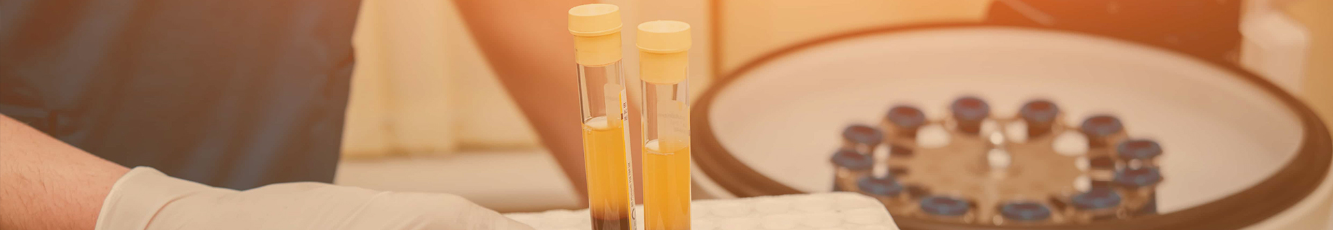
PRP is a concentrated autologous full platelet. Compared with normal platelets, its concentration is 4-5 times higher. High concentration platelets with coordinase and calcium ions The flocculent gels formed are called richer platelet gels, which can participate in the establishment of the cell branch architecture. PRP contains many types of protein and cytokines, such as common platelet derivative growth factor (PDGF), vascular endothelial growth factor (VEGF), and fibrin. The alpha particles released after the above growth factors can play a repair role, thereby promoting bone healing and reconstruction of vascular reconstruction. PRP contains protein that promotes cartilage cell cell adhesion, thereby ensuring tissue repair. PRP is separated from autologous blood, and its safety has been confirmed by theoretical and related animal experiments. PRP is not only safe, but also has the characteristics of regenerative biological characteristics, which has a significant repair effect on cartilage and tissue damage.
2) Proliferation mechanism of cartilage cells
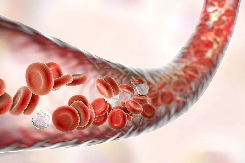
VEGF and Fiber cell growth factor (FGF) are related to the reconstruction of vascular reconstruction. Under the action of VEGF, the proliferation of vascular endothelial cells can help new blood vessel formation and the improvement of blood transportation of the affected area, thereby promoting tissue healing. FGF can also regulate cells to accelerate the proliferation of vascular endothelial cells by regulating cells. Hepatocyte growth factor (HGF) can activate nuclear factors-XB (NF-XB), leukocyte (IL) -1 to inhibit the inflammatory response of chondrocytes. The internal fiber protein molecular content of PRP has a high content. It can form a 3D grid fiber under activation of coordinase and calcium ion. Therefore, PRP can also be called a platelet gel. PRP can not only promote the repair of cartilage tissue, but also provide an attached bracket for cartilage anterior cell cells and promote its differentiation, which will help the formation of transparent cartilage matrix. It can be seen that PRP not only helps the reconstruction of the cartilage and vascular region and the synthesis of cartilage fiber brackets, but also promotes the adhesion and migration of the cartilage cells of the cartilage, and then repair the damage of the cartilage tissue.
3) PRP experimental research on the half moon board repair
Some scholars chose rabbits as experiments, and after the half moon board defects were made in both knees, the rabbits were executed at 4 weeks and 8 weeks, and their pathological performance was analyzed. The study found that at 4 weeks, the control group half moon plate was composed of connective tissue, which can be manifested as severe fibrosis; and the PRP treatment group’s half moon plate structure showed normal, and the junction tissue had obvious repairs. Organization composition. At 8 weeks, the control group was full of fibrous tissue, and the cartilage of the half -moon plate was not formed. The PRP therapy group was rich in fiber in the half moon plate, which increased significantly. At the same time, the half moon plate tissue manifests in moderate fibrosis, and even partial healing is present. Another study has found that fibrin in pre-processing PRP can form a mesh stent consisting of polystumin-hydroxylcetic acid cluster. If the PRP is centrifuged, and the half moon cartilage chondrocyte is combined to cultivate 7D into the nude mice of the experimental group, the fluorescent microscope examination: The cartilage cells after the seedlings are sowing can be evenly stuck and spread all over the bracket. After PRP is treated, the number of cartilage cells has increased significantly. The results of scanning electron microscopy show that on the processed PRP brackets, cartilage cells can be connected to the fiber protein network after 24H and 7D. Among the 16 cases of processing the PRP bracket group mice, 6 cases were completely healed, 9 cases were incomplete, and 1 case did not heal, while the control group mice did not heal. It can be seen that after the PRP is processed, human joint cartilage cells have specific cell adhesion capabilities, which can enhance the half moon board healing ability. In vitro and in vitro test research, compared with the simple PRP gel group, the PRP-osteoma matrix cell gel treatment group has a higher degree of differentiation. bone. There are studies on the combined PRP processing of the Jackie Daping model of the rabbit cartilage, and keep the cartilage defective area passive movement. The results of the immunohistochemistry and the fault scanning test have confirmed that its tissue repair score is high. The result suggests that PRP plays an important role in cartilage repair, and the combined application of the combined application of the combined application of the bone marrow mesenchymal stem cells and the bracket material is better.
(The contents of this article are reprinted, and we do not provide any express or implied guarantee for the accuracy, reliability or completeness of the contents contained in this article, and are not responsible for the opinions of this article, please understand.)
Post time: May-11-2023